
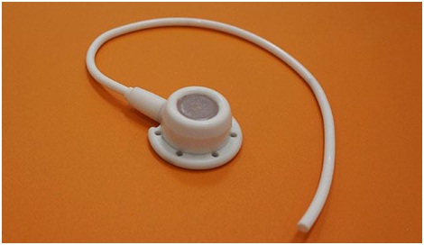
A chemo port is a small-sized vein-access device that has implantable reservoir with a thin silicon tube that attaches to a vein. The main advantage of this device is that chemotherapy medications can be delivered directly into the port rather than a vein, eliminating the need for needle sticks. Patients who receive chemotherapy choose to have a port implanted if recommended by their treatment team.
It is not always required to remove benign tumours or to treat them further. A significant volume of ongoing research studies about treatment methods is done with cancerous tumours. In multicenter clinical trials, novel chemotherapy drugs are being tested and researchers are trying to identify molecular targets for cancerous growths to help in designing the drugs aimed at curing such cancer. Techniques like metabolic imaging PET (Positron Emission Tomography) scanning is opening new avenues to evaluate tumour response to chemotherapy and to identify the aggressive tumours.
A chemo port is placed centrally in the upper chest under the skin near a large vein. This can be a good alternative to an intravenous (IV) catheter that is superficially placed in an arm or hand vein due to which a suitable IV site can sometimes be difficult to find. A chemo port is easily accessible by a patient’s treatment team, a port can provide a safer and more efficient medication delivery process than an IV. A port site is prepared using a sterile technique, that means all surfaces are free of microorganisms reducing the risk of infection to great extent, whereas an IV site is prepared with a clean but nonsterile technique. A port can be used for also for drawing blood for lab testing, delivering fluids and transfusions and injecting dye for PET and CT scans.
For the procedure, the patient is transferred to the procedure bed in the angiography room. Then the patient's one side of neck and chest position is set for the procedure depending on the accessibility to the blood vessels. After this sterilization of the body area where port placement is to be done and other parts of the body are covered with a sterile sheet. After that doctor uses the medication to numb the area of neck access and inserts a needle to access the internal jugular vein in the neck of the patient with the help of ultrasonic guidance. Once the access is achieved the doctor will introduce a guidewire into the large vein in the chest called SVC (Superior Vena Cava), where catheter tip will be eventually placed. The X-ray machine is used to ensure the proper placement of the wire.
Next, the doctor will numb the site for port placement and the areas of the chest that will be tunnelled for the catheter to be secured under. Once the patient is numb, the doctor will use a scalpel to create a 3 cm long skin incision and then create a pocket for the port underneath it using a blunt dissector. The only sensation patient feels at this time is pressure. If at any time patient would feel pain or discomfort and informs the doctor about it, then more numbing medication is given by the doctor.
Next, the doctor will use a tunnelling device to create a path under the skin from the port site to the access site on the neck. He will then tunnel the catheter tip under the tunnel with the port attached to other ends. After this the doctor will place a sheath, that is the device to help catheter placement, over the wire and into the patient’s vein. At this point, the doctor will ask the patient to take a deep breath and hold it, and threads the catheter tip over the wire and briskly advances it through the sheath and to SVC. Once the catheter tip is in place, the doctor will use imaging to confirm and document its proper placement. The doctor will then close the port site and the access point in the neck with sutures and skin glue. The function of the port will be verified with aspiration and flushing of saline and heparin. After this, the port will be ready to use. The doctor will finish the procedure by cleaning the patient’s neck and chest.
Though a port produces a visible, quarter-sized bump under the skin, it is easily coverable with regular clothing. It can remain in place until it is necessary, for many weeks, unlike an IV catheter, which must be reinserted for each treatment session, for many months or even years. It can be easily removed through a relatively simple outpatient procedure when it is no longer needed. This way the port helps to streamline the delivery of chemotherapy medications.
The port removal process will also be performed at the angiography suite under sterile conditions. It will involve the use of numbing indication and reopening of the port pocket to remove the device. Post which the pocket is reclosed with suture and skin glue.